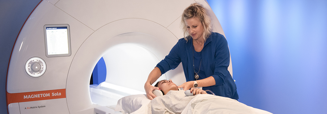
Beaumont is committed to providing high quality MRI imaging services with a focus on patient and family centered care. We offer:
- ease of scheduling by contacting our access center
- many convenient locations for MRI imaging in South East Michigan.
- board certified Beaumont radiologists to interpret your results.
- committed to providing the most up-to-date imaging technology
- highly trained technologists to ensure your comfort and safety
- an integrated electronic medical records system that allows your Beaumont Health care team to promptly see your images.
What is Magnetic Resonance Imaging?
Magnetic resonance imaging (MRI) is a medical imaging technique that uses a magnetic field and computer-generated radio waves to create detailed images of the organs and tissues in your body.
Most MRI machines are large, tube-shaped magnets. When you lie inside an MRI machine, the magnetic field temporarily realigns water molecules in your body. Radio waves cause these aligned atoms to produce faint signals, which are used to create cross-sectional MRI images — like slices in a loaf of bread. A patient will not be able to feel any of these changes taking place inside the body.
How does an MRI scan work?
- The patient is placed on a table within an active magnetic field. Pulses of radio waves (a loud knocking noise) are created within the scanner.
- Your body is made of tiny bits of matter called atoms; at the center of each atom is a nucleus. The radio waves move the nuclei of the atoms in your body out of their normal position.
- As the nuclei realign into proper position, they send out radio signals.
- The signals are received by a computer that converts them into an image.
- The images are interpreted by a board certified radiologist and the results are sent to your ordering physician.
What to expect at your MRI
An MRI can help you and your doctor better understand what may or may not be occurring and aid in diagnosis. Learn more about the test you are having.
Patient Preparation
Before an MRI exam, eat normally and continue to take your usual medications, unless otherwise instructed. Upon scheduling, you will be given detailed instructions regarding any kind of prep required, notes on whether you may eat or drink prior to the exam, and whether you need to discontinue medication. You will be asked to change into a gown and to remove things that might affect the magnetic imaging or cause a safety concern. Lockers are provided at each scanner to hold valuables while the exam is taking place.
Some examples of items that will need to be removed:
- Jewelry
- Hairpins
- Eyeglasses
- Watches
- Wigs
- Dentures
- Hearing aids
- Underwire bras
- Medication Patches
- Cell phones, keys, loose change, pocket knives, credit cards
- External Pumps/Monitors
Note: All wearable monitors must be removed prior to imaging. Consider waiting to apply a new patch until after your MRI if you are close to finishing with your current patch and having new monitor available to use after your test. Contact the manufacturer of your monitor for additional details.
Infants and children under the age of 12 are not permitted to accompany their parent/guardian during any imaging exam or procedure.
What to expect
The MRI machine looks like a long narrow tube that is open at both ends. You will lie down on a movable table that slides into the opening of the tube. A technologist monitors you from another room. You can talk with the person by microphone.
If you have a fear of enclosed spaces, you will want to notify your physician. They can prescribe you medication to help you feel less anxious. Most people get through the exam without difficulty. Anesthesia is offered at multiple sites as well for those who require more extensive sedation.
During the MRI scan, the internal part of the magnet produces repetitive tapping, thumping and other noises. You will be given earplugs and headphones to help block the noise. For some exams, music can be played or a video can be watched to help distract from the noise and make patients less anxious.
In some cases, a contrast material, called gadolinium, will be injected through an intravenous (IV) line into a vein in your hand or arm. The contrast material enhances certain details and provides additional information to aid the Radiologists in making a diagnosis.
Standard Bore
- 60cm Bore diameter
- fits most patients comfortably
- located at our Royal Oak and Troy
Wide Bore
- 70 cm Bore diameter
- designed to help reduce claustrophobia
- shorter front to back and wider side to side than traditional MRI's
- located in Dearborn, Farmington Hills, Grosse Pointe, Lake Orion, Lennox, Livonia, Royal Oak, Taylor, Trenton, Troy and Wayne
Bore height is measured from the top to bottom of the opening.
Risks
Because MRI uses powerful magnets, the presence of metal in your body can be a safety hazard. Even if not attracted to the magnet, metal objects can distort the MRI image. Before having an MRI, you'll complete a questionnaire that includes questions regarding any metal or electronic devices in your body. It is very important to answer truthfully and accurately. You may be asked to bring implant information cards with you to the appointment to show the Technologist prior to your scan.
Unless the device you have is certified as MRI safe/conditional, you may not be able to have an MRI. Devices include:
- Metallic joint prostheses
- Artificial heart valves
- Implanted drug infusion pumps
- Implanted nerve stimulators
- A pacemaker or defibrillator
- Metal clips, stents, filters, coils
- Metal pins, screws, plates, or surgical staples
- Cochlear implants
- A bullet, shrapnel, or any other type of metal fragment