Utilizing serum/plasma and peripheral monocytes for AD diagnosis and understanding pathogenesis mechanism - The presence of higher level of plasma matrix proteins such as albumin alpha2-macroglobulin, lipoprotein, etc., hinders the accuracy of immunoassays
to detect the amount of Aβ in serum/plasma samples. Evidence suggests that plasma Aβ42 and Aβ40 levels are associated with the levels of CSF Aβ42/Aβ40 and cerebral amyloid deposition. Regardless of the amount of monomeric
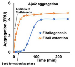
Aβ42 or Aβ40, we aim to determine the potential Aβ aggregates in serum using seed extension methods to diagnose AD using noninvasive methods.
 Fig: Aβ aggregation progresses via two steps, nucleation and fibril extension. During nucleation, smaller aggregates are formed by assembly of multiple monomeric species. During the extension phase, monomers are attached to existing oligomers that extend the fibrils. The nucleation (seed formation/lag) phase can be omitted by adding preformed oligomers/fibrils/seeds in the reaction mixture.
It has been shown that significant portion of brain macrophages are migrated from peripheral monocytes upon acute or chronic brain injuries. We aim to identify subsets of peripheral monocytes that potentially participate in inflammatory response and migrate to brain in AD patients.