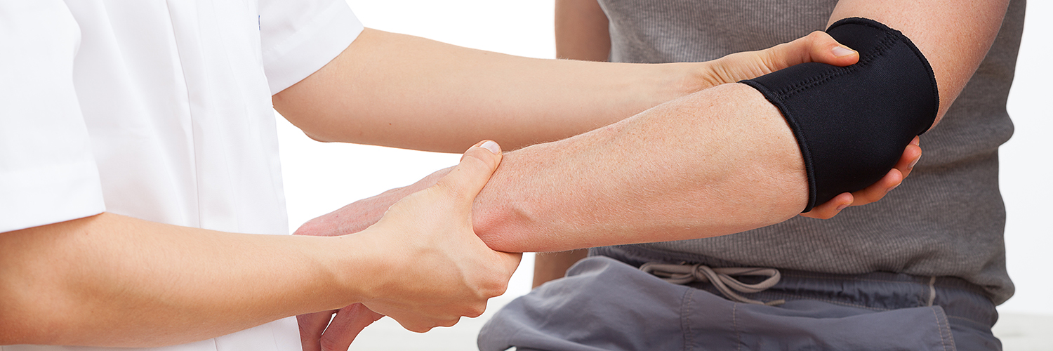
 Lateral epicondylitis, also known as tennis elbow, is characterized by pain on the outside (lateral side) of the elbow. The pain is caused by damage to the tendons that bend the wrist backward away from the palm. A tendon is a tough cord of tissue that connects muscles to bones. The tendon most likely involved in tennis elbow is called the extensor carpi radialis brevis, and this condition is usually diagnosed in both men and women between the ages of 30 years to 50 years.
Causes of Tennis Elbow
Tennis elbow, as the name implies, often is caused by the force of the tennis racket hitting balls in the backhand position. The forearm muscles, which attach to the outside of the elbow, may become sore from excessive strain. When making a backhand stroke in tennis, the tendons that roll over the end of the elbow can become damaged. Tennis elbow may be caused by the following:
- improper backhand stroke
- weak shoulder and wrist muscles
- using a racket that is too tightly strung or too short, such as one that is meant for racquetball or squash
- hitting the ball off center on the racket or hitting heavy, wet balls
However, many people who suffer from lateral epicondylitis do not play tennis. The condition is caused by any repetitive movement. Other causes of tennis elbow include:
- painting with a brush or roller
- operating a chain saw
- frequent use of other hand tools on a continuous basis
- using repeated hand motions in various professions, such as meat cutters, musicians, dentists, and carpenters
Symptoms of Tennis Elbow
The following are the most common symptoms of tennis elbow. However, each individual may experience symptoms differently.
Initially, the pain may be felt along the outside of the forearm and elbow. The pain may increase down to the wrist, even at rest, if the person continues the activity that causes the condition. Pain may also persist when the arm and hand are placed palm-down on a table and the person tries to raise the hand against resistance, or with lifting and gripping small objects, such as a coffee cup.
The symptoms of tennis elbow may resemble other medical problems or conditions. Always consult your doctor for a diagnosis.
Diagnosis of Tennis Elbow
The diagnosis of tennis elbow usually can be made based on a physical examination. However, in some cases, an X-ray, magnetic resonance imaging (MRI), and electromyography (EMG) of the elbow are necessary.
Treatment for Tennis Elbow
Specific treatment for tennis elbow will be determined by your doctor based on:
- your age, overall health, and medical history
- extent of the condition
- your tolerance for specific medications, procedures, and therapies
- expectation for the course of the condition
- your opinion or preference
Treatment for tennis elbow includes stopping the activity that produces the symptoms. It is important to avoid the movement that caused the injury in the first place. Treatment may include:
- ice pack application (to reduce inflammation)
- strengthening exercises
- anti-inflammatory medications
- bracing
- corticosteroid injections
- surgery
Advanced Treatment: Tenex Tenotomy
The Tenex tenotomy procedure is a non-surgical procedure used to treat chronic pain associated with tendinitis/tendinopathy and plantar fasciitis/fasciosis. The minimally invasive technique can reduce tendon pain by breaking down and removing damaged tissues with high-frequency ultrasound energy. The procedure is commonly used to treat tendinitis/tendinopathy of the elbow, hip, knee, shoulder, ankle, and plantar fascia. The procedure is performed using local anesthetic and ultrasound guidance which makes it extremely safe. The procedure is minimally invasive and allows patients to return to normal activities faster than surgery.
The procedure is performed though a small skin puncture (2-3mm) and the device is advanced to the diseased tendon or plantar fascia using ultrasound guidance. The device then removes the diseased tissue and stimulates your bodies normal healing response. The device is then removed and small bandage is applied. Patients go home shortly after the procedure and typically have a short course (3-7 days) of relative immobilization in a sling or walking boot.